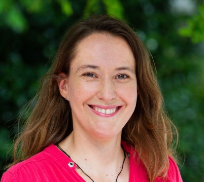
Here we profile research led by Dr Julie Choisne from the Auckland Bioengineering Institute that is helping to bring computational modelling into clinical practice.
Childhood conditions such as developmental hip abnormalities, cerebral palsy and slipped capital femoral epiphysis (a hip condition that occurs in teens and pre-teens who are still growing) can lead to complex hip and knee deformities. Due to the variability in paediatric conditions, outcomes of surgical interventions are variable, making it difficult to quantify the effectiveness of individual treatments or know which are most effective for which child.
To help solve this problem, Dr Julie Choisne from the Auckland Bioengineering Institute used funding from an HRC Emerging Researcher First Grant to obtain more than 330 CT scans of children aged from 4 to 18 years and manually segment the bones in the lower extremity. From here, Dr Choisne and her team created the world’s first 3D bone growth chart of the pelvis, femur and tibia/fibula for children aged 4 to 18 years old.
This new tool can serve as a reference to understand and compare bone measurements in children with atypical bone development. These measurements are freely available on an opensource database accessible to the clinical and research community.
Dr Choisne’s team have also created a world-first computer model that can accurately predict a child’s skeleton based on their age, sex, height and mass. This model can be used worldwide and is now being implemented in all gait clinics in Australia and New Zealand for use on children with movement disorders.
“This model will reduce processing times for 3D gait analysis by automatically computing each child's joint range of motion and moments during gait. Moreover, we can now create a database of gait analysis that can train AI models to help physiotherapists, orthopaedic surgeons, orthotists etc in their assessment and treatment decision-making,” says Dr Choisne.
