Microvascular abnormalities identified using retinal imaging are providing valuable clues as to what lies behind schizophrenia.
The research, which was a collaboration between the HRC-funded Dunedin Multidisciplinary Health and Development Research Unit (DMHDRU) and the Singapore Eye Research Institute (SERI), was carried out as part of the age 38 follow-up for the Dunedin longitudinal study. The Dunedin Study has been following a cohort of 1000 people born in the city in 1972-3.
Director of the University of Otago-based unit Professor Richie Poulton says retinal imaging is relatively new technology and this is the first time they have used it as part of a follow-up.
The opportunity came as the result of visiting other Singaporean collaborators who introduced him to Dr Tien Wong, a world authority on retinal imaging, who heads up SERI.
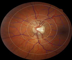
Professor Poulton says that by taking a picture of the back of the retina they can examine the micro vessels, effectively giving them a window into many facets of an individual's health.
In the case of schizophrenia, they are particularly interested in identifying anyone with wider retinal venules, which tend to reflect insufficient brain oxygen supply.
"It is effectively an opportunity to look inside them without having to cut them open. So the non-invasive and relatively practical nature of the assessment is a real boon to researchers.”
"It hasn't been used much and certainly hasn't been used much outside ophthalmology. It is about correlates of bad health that you can access through the eyes, such as diabetes."
But having the vast Dunedin Study database means they have measurements of all sorts of health areas, including psychiatry and mental health.
Professor Poulton says they were told to expect about 10 per cent data loss because it is quite difficult to get clear images. However, they managed to get a 98 per cent success rate in terms of clear images.
"The Dunedin Study team were able to show the association between widened venules and schizophrenia. But we were also able to show that it wasn't just a function of other known risk factors of having wide venules, which we were able to control for."
Professor Poulton says the study is also important at the method level.
"This is an interesting way of approaching and understanding the pathogenesis and development of this incredibly burdensome problem of schizophrenia.”
"Despite 100 years of research we still don't have a really good handle on why it is that some people develop this problem. There are a lot of theories – and you need really good tools to test those theories. This is evidence that this particular tool may be very useful for understanding schizophrenia," he says.
"This is also proof that these longitudinal studies shouldn't just be about epidemiological studies of pathways and prevalence. They can be wonderful resources for beginning to dig down deep into biology."
Professor Poulton says retinal imaging will continue to be part of future assessments. These will include tracking changes in cognitive function and relating that to important areas such as mild cognitive impairment, which is a precursor to neurodegenerative disorders and dementia.